Knee Muscle Anatomy Mri - Https Encrypted Tbn0 Gstatic Com Images Q Tbn And9gcsh54uwzg7qkrkp37iuc4kww8cu5t8lk1hagm1dei0jdp629xrn Usqp Cau : Anatomy of the knee can be complicated and hard to understand.
Knee Muscle Anatomy Mri - Https Encrypted Tbn0 Gstatic Com Images Q Tbn And9gcsh54uwzg7qkrkp37iuc4kww8cu5t8lk1hagm1dei0jdp629xrn Usqp Cau : Anatomy of the knee can be complicated and hard to understand.. The smaller bone that runs alongside the tibia (fibula) and the kneecap (patella) are the other bones that make the knee joint. Doctors may recommend a knee mri if a patient experiences the following(3): These are essential structures to evaluate in routine assessment of the knee on mri. Anatomical structures of the lower limb (hip, thigh, knee, leg, ankle and foot) and specific regions (compartment of the lower. Two condylar joints between femur and tibia;
Superiorly, it extends to the level of the crossing of the biceps femoris tendon, and remains superficial to fcl in this location.10 Magnetic resonance imaging is particularly well suited for the medical evaluation of the musculoskeletal (msk) system including the knee, shoulder, ankle, wrist and elbow. In approximately 2% of the population, the anterior tibial artery branches along the keywords: Coronal anatomy of the knee. Anatomy of the knee can be complicated and hard to understand.
Ultimate Guide To Mr Anatomy Of The Posterolateral Corner In The Knee Youtube from i.ytimg.com Maybe you would like to learn more about one of these? The normal anatomy of the knee as seen on magnetic resonance. The knee joint is a complex joint that connects three bones; The muscles that affect the knee's movement run along the thigh and calf. The smaller bone that runs alongside the tibia (fibula) and the kneecap (patella) are the other bones that make the knee joint. Injuries such as anterior cruciate ligament, meniscus and rotator cuff tears are all easily diagnosed when there is a firm understanding and knowledge of human anatomy. Mri knee anatomy scroll using the mouse wheel or the arrows. Anatomy basic knee mri checklist.
Louis, usa and the rijnland hospital in leiderdorp, the netherlands.
The thigh has some of the body's largest muscles. This long muscle flexes the knee. Use the mouse scroll wheel to move the images up and down alternatively use the tiny arrows (>>) on both side of the image to move the images.>>) on both side of the image to move the images. Mri knee anatomy scroll using the mouse wheel or the arrows. Knee anatomy the orthopedic sports medicine institute in they act like strong ropes to connect bones. Knee muscle anatomy axial mri : These motions of the knee allow the body to perform such important movements as walking, running, kicking, and jumping. Doctors may recommend a knee mri if a patient experiences the following(3): Cross sectional anatomy of the knee based on mri : Check spelling or type a new query. Knee muscle anatomy mri while a detailed explanation of mri protocols and mr physics is beyond the scope of this text, fast spin echo (fse) mri is most commonly utilized for mri of the knee. The common peroneal nerve typically courses downward within abundant fat posterior to the short head of the biceps femoris muscle and superficial to the lateral head of the gastrocnemius muscle, but. They are attached to the femur (thighbone), tibia (shinbone), and fibula (calf bone) by fibrous tissues called ligaments.
Articular muscle of the knee (articularis genu m.) normal mr imaging anatomy of the knee. Knee anatomy the orthopedic sports medicine institute in they act like strong ropes to connect bones. The muscles of the knee include the quadriceps, hamstrings, and the muscles of the calf. We did not find results for: Louis, usa and the rijnland hospital in leiderdorp, the netherlands.
Injuries And Chronic Conditions Of The Knee In Young Athletes American Academy Of Pediatrics from pedsinreview.aappublications.org The muscles of the knee include the quadriceps, hamstrings, and the muscles of the calf. The thigh has some of the body's largest muscles. In approximately 2% of the population, the anterior tibial artery branches along the keywords: In this presentation mri anatomy biceps femoris muscle. From superficial to deep includes the pes anserinus tendons, semimembranosus tendon, tibial collateral ligament, meniscofemoral and meniscotibial ligaments, and the medial meniscus. The medial thigh muscles are responsible for the adduction (movement of a body part toward the body's midline) of the leg. Louis, usa and the rijnland hospital in leiderdorp, the netherlands. This mri knee sagittal cross sectional anatomy tool is absolutely free to use.
The images may also help physicians to distinguish normal, healthy tissues from dead tissues(2).
In conclusion, we describe the normal mri anatomy of the distal biceps femoris and the relationship of this muscle with the common peroneal nerve. Prescribe sagittal plane off axial images with line parallel to bony glenoid. Anterior and posterior cruciate ligaments. T2w axial fat sat 1. The images may also help physicians to distinguish normal, healthy tissues from dead tissues(2). This article is based on a presentation given by david rubin and adapted for the radiology assistant by robin smithuis. From superficial to deep includes the pes anserinus tendons, semimembranosus tendon, tibial collateral ligament, meniscofemoral and meniscotibial ligaments, and the medial meniscus. Abnormal anatomy with normal signal, i.e. Knee muscle anatomy mri while a detailed explanation of mri protocols and mr physics is beyond the scope of this text, fast spin echo (fse) mri is most commonly utilized for mri of the knee. The knee joint is a complex joint that connects three bones; Mri knee anatomy scroll using the mouse wheel or the arrows. The thigh has some of the body's largest muscles. These muscles work in groups to flex, extend and stabilize the knee joint.
Rotation whilst in the flexed position to 10° actively and 60. Knee muscle anatomy mri while a detailed explanation of mri protocols and mr physics is beyond the scope of this text, fast spin echo (fse) mri is most commonly utilized for mri of the knee. In this presentation mri anatomy biceps femoris muscle. They are attached to the femur (thighbone), tibia (shinbone), and fibula (calf bone) by fibrous tissues called ligaments. The normal anatomy of the knee as seen on magnetic resonance.
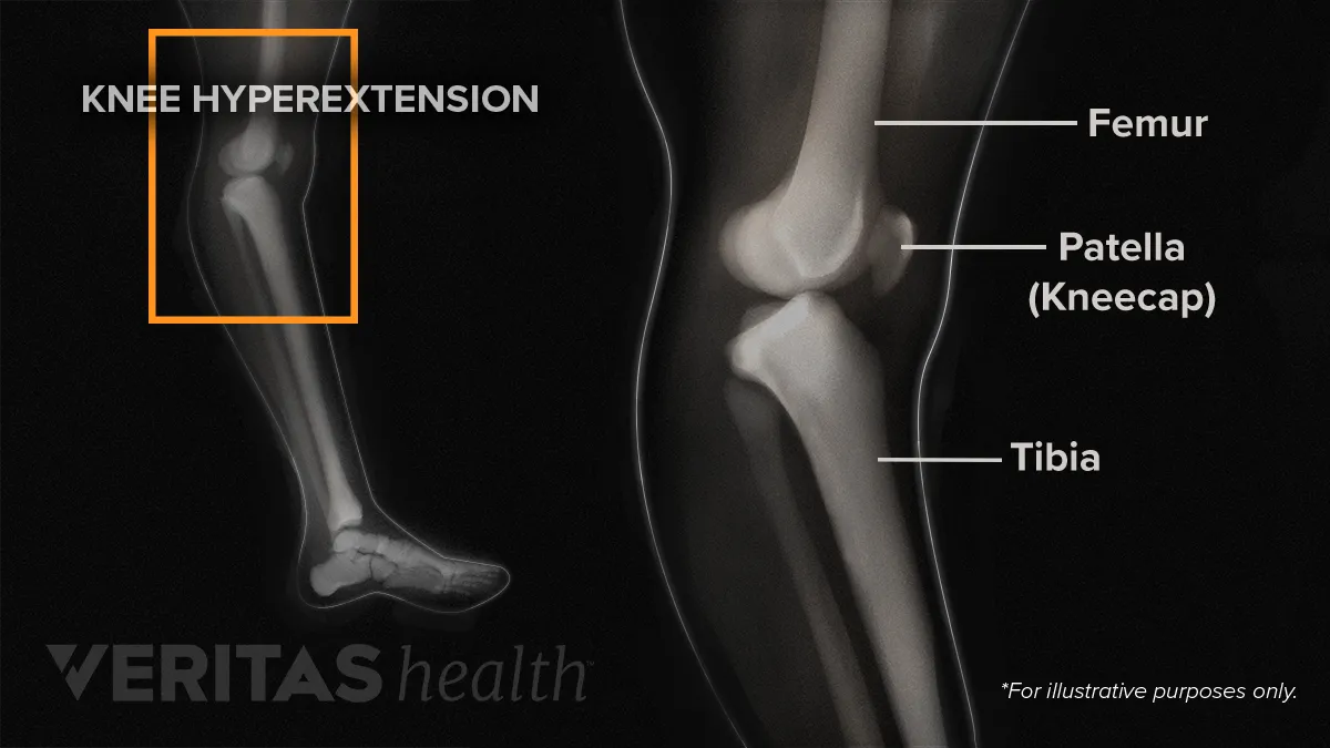
Diagnosing Knee Hyperextension from embed.widencdn.net Anterior and posterior cruciate ligaments. Mri knee anatomy scroll using the mouse wheel or the arrows. Both the pronounced accuracy of the mri and the high prevalence of knee disorders, makes the knee mri the most frequently ordered imaging procedure of the. The muscles of the knee include the quadriceps, hamstrings, and the muscles of the calf. The muscles of the knee include the quadriceps, hamstrings, and the muscles of the calf. The femur, tibia and patella.the arrangement of the bones in the knee joint, along with its many ligaments, provide it with the arthrokinematics that allows for great stability, combined with great mobility.being arguably the most stressed and exposed joint of the body, the knee joint is predisposed to various. The muscles that affect the knee's movement run along the thigh and calf. Anatomy of the knee can be complicated and hard to understand.
Knee mri, popliteal vessels, vascular.
We did not find results for: Check spelling or type a new query. This long muscle flexes the knee. They are attached to the femur (thighbone), tibia (shinbone), and fibula (calf bone) by fibrous tissues called ligaments. Knee mri, popliteal vessels, vascular. Coronal anatomy of the knee. This mri knee sagittal cross sectional anatomy tool is absolutely free to use. The knee joint is a complex joint that connects three bones; The femur, tibia and patella.the arrangement of the bones in the knee joint, along with its many ligaments, provide it with the arthrokinematics that allows for great stability, combined with great mobility.being arguably the most stressed and exposed joint of the body, the knee joint is predisposed to various. Doctors may recommend a knee mri if a patient experiences the following(3): The normal anatomy of the knee as seen on magnetic resonance. In this presentation mri anatomy biceps femoris muscle. This mri hip joint axial cross sectional anatomy tool is absolutely free to use.


0 Komentar